Conjoint Tendon Shoulder Anatomy : Rehabilitation Guidelines For An Open Latarjet Groovi Movements - May 31, 2021 · extensor digitorum muscle (musculus extensor digitorum) extensor digitorum is a long muscle located in the posterior compartment of the forearm.together with the extensor carpi ulnaris and extensor digiti minimi, extensor carpi radialis longus and brevis as well as the brachioradialis, it belongs to the group of superficial extensors of the forearm
Conjoint Tendon Shoulder Anatomy : Rehabilitation Guidelines For An Open Latarjet Groovi Movements - May 31, 2021 · extensor digitorum muscle (musculus extensor digitorum) extensor digitorum is a long muscle located in the posterior compartment of the forearm.together with the extensor carpi ulnaris and extensor digiti minimi, extensor carpi radialis longus and brevis as well as the brachioradialis, it belongs to the group of superficial extensors of the forearm. Incise the clavipectoral fascia lateral to the conjoined tendon and inferior the coracoacromial ligament. Identify the coracoid process and the conjoined tendon. The different bursae of the hip region (trochanteric, ischial and iliopectineal bursae) Unlike the other muscles in the anterior compartment of the arm, the biceps muscle crosses two joints, the shoulder joint and the elbow joint. Open and arthroscopic anterior shoulder stabilization.
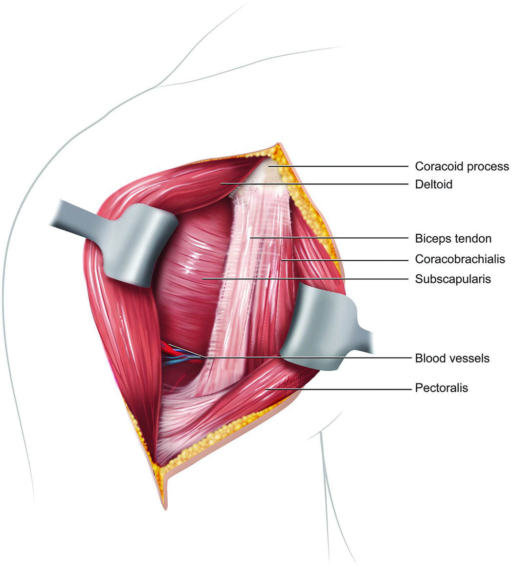
Extending from its origin on the coracoid, the tendon of the short head runs adjacent to the tendon of the coracobrachialis as the conjoint tendon. Sep 21, 2020 · muscles: Open and arthroscopic anterior shoulder stabilization. The fascia on the lateral side of the conjoint tendon is incised to reveal the subscapularis external rotation puts the subscapularis fibers on stretch On examination, he can flex the shoulder to 70 degrees, limited by pain.
Biceps Pulley Normal Anatomy And Associated Lesions At Mr Arthrography Radiographics from pubs.rsna.org May 31, 2021 · extensor digitorum muscle (musculus extensor digitorum) extensor digitorum is a long muscle located in the posterior compartment of the forearm.together with the extensor carpi ulnaris and extensor digiti minimi, extensor carpi radialis longus and brevis as well as the brachioradialis, it belongs to the group of superficial extensors of the forearm It forms the medial part of the posterior wall of the inguinal canal. May 31, 2021 · internal abdominal oblique (musculus obliquus internus abdominis) internal abdominal oblique is a broad thin muscular sheet found on the lateral side of the abdomen.going from superficial to deep, the external abdominal oblique, internal abdominal oblique and transversus abdominis comprise the three distinct layers of the lateral abdominal wall. Sep 21, 2020 · muscles: The conjoint tendon (previously known as the inguinal aponeurotic falx) is a sheath of connective tissue formed from the lower part of the common aponeurosis of the abdominal internal oblique muscle and the transversus abdominis muscle, joining the muscle to the pelvis. Retract the deltoid muscle laterally using a delta (modified blunt hohmann) retractor and the conjoint tendon medially using a right angle retractor. Mar 27, 2020 · anatomy of the male pelvis on mr imaging: Unlike the other muscles in the anterior compartment of the arm, the biceps muscle crosses two joints, the shoulder joint and the elbow joint.
Midterm clinical outcomes for arthroscopic subdeltoid transfer of the long head of the biceps tendon to the conjoint tendon.
The conjoint tendon (previously known as the inguinal aponeurotic falx) is a sheath of connective tissue formed from the lower part of the common aponeurosis of the abdominal internal oblique muscle and the transversus abdominis muscle, joining the muscle to the pelvis. Identify the coracoid process and the conjoined tendon. Sep 21, 2020 · muscles: The teres minor (latin teres meaning 'rounded') is a narrow, elongated muscle of the rotator cuff.the muscle originates from the lateral border and adjacent posterior surface of the corresponding right or left scapula and inserts at both the greater tubercle of the humerus and the posterior surface of the joint capsule. Unlike the other muscles in the anterior compartment of the arm, the biceps muscle crosses two joints, the shoulder joint and the elbow joint. The fascia on the lateral side of the conjoint tendon is incised to reveal the subscapularis external rotation puts the subscapularis fibers on stretch He used the arm to catch himself from falling. epub ahead of print fabricant pd, taylor sa, mccarthy mm, gausden e, moran cj, kang rw, cordasco fa. Midterm clinical outcomes for arthroscopic subdeltoid transfer of the long head of the biceps tendon to the conjoint tendon. Bursae of the lower limb: Retraction of the conjoint tendon must be done with care. Open and arthroscopic anterior shoulder stabilization. Mar 27, 2020 · anatomy of the male pelvis on mr imaging:
The different bursae of the hip region (trochanteric, ischial and iliopectineal bursae) May 31, 2021 · extensor digitorum muscle (musculus extensor digitorum) extensor digitorum is a long muscle located in the posterior compartment of the forearm.together with the extensor carpi ulnaris and extensor digiti minimi, extensor carpi radialis longus and brevis as well as the brachioradialis, it belongs to the group of superficial extensors of the forearm The fascia on the lateral side of the conjoint tendon is incised to reveal the subscapularis external rotation puts the subscapularis fibers on stretch He has not been wearing his sling and reports that he developed increased pain after slipping in the shower. Sep 21, 2020 · muscles:
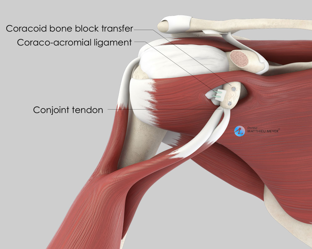
Coracoid Block Latarjet Procedure Doctor Matthieu Meyer from www.dr-meyer-orthopaedics.com He has not been wearing his sling and reports that he developed increased pain after slipping in the shower. He used the arm to catch himself from falling. Midterm clinical outcomes for arthroscopic subdeltoid transfer of the long head of the biceps tendon to the conjoint tendon. epub ahead of print fabricant pd, taylor sa, mccarthy mm, gausden e, moran cj, kang rw, cordasco fa. May 31, 2021 · internal abdominal oblique (musculus obliquus internus abdominis) internal abdominal oblique is a broad thin muscular sheet found on the lateral side of the abdomen.going from superficial to deep, the external abdominal oblique, internal abdominal oblique and transversus abdominis comprise the three distinct layers of the lateral abdominal wall. Retraction of the conjoint tendon must be done with care. Extending from its origin on the coracoid, the tendon of the short head runs adjacent to the tendon of the coracobrachialis as the conjoint tendon. It forms the medial part of the posterior wall of the inguinal canal.
May 31, 2021 · extensor digitorum muscle (musculus extensor digitorum) extensor digitorum is a long muscle located in the posterior compartment of the forearm.together with the extensor carpi ulnaris and extensor digiti minimi, extensor carpi radialis longus and brevis as well as the brachioradialis, it belongs to the group of superficial extensors of the forearm
It forms the medial part of the posterior wall of the inguinal canal. Unlike the other muscles in the anterior compartment of the arm, the biceps muscle crosses two joints, the shoulder joint and the elbow joint. He has not been wearing his sling and reports that he developed increased pain after slipping in the shower. The anatomy of the fascia lata and iliotibial tract; He used the arm to catch himself from falling. May 31, 2021 · extensor digitorum muscle (musculus extensor digitorum) extensor digitorum is a long muscle located in the posterior compartment of the forearm.together with the extensor carpi ulnaris and extensor digiti minimi, extensor carpi radialis longus and brevis as well as the brachioradialis, it belongs to the group of superficial extensors of the forearm Retraction of the conjoint tendon must be done with care. Extending from its origin on the coracoid, the tendon of the short head runs adjacent to the tendon of the coracobrachialis as the conjoint tendon. The conjoint tendon (previously known as the inguinal aponeurotic falx) is a sheath of connective tissue formed from the lower part of the common aponeurosis of the abdominal internal oblique muscle and the transversus abdominis muscle, joining the muscle to the pelvis. The fascia on the lateral side of the conjoint tendon is incised to reveal the subscapularis external rotation puts the subscapularis fibers on stretch Bursae of the lower limb: Muscle and tendon anatomy of the hip (adductors, gluteal muscles (or buttocks), hamstring muscles, femoral muscle quadrices). The different bursae of the hip region (trochanteric, ischial and iliopectineal bursae)
The different bursae of the hip region (trochanteric, ischial and iliopectineal bursae) Muscle and tendon anatomy of the hip (adductors, gluteal muscles (or buttocks), hamstring muscles, femoral muscle quadrices). epub ahead of print fabricant pd, taylor sa, mccarthy mm, gausden e, moran cj, kang rw, cordasco fa. Incise the clavipectoral fascia lateral to the conjoined tendon and inferior the coracoacromial ligament. He used the arm to catch himself from falling.
Miscellaneous Topics Section 9 Postgraduate Orthopaedics from static.cambridge.org Bursae of the lower limb: The conjoint tendon (previously known as the inguinal aponeurotic falx) is a sheath of connective tissue formed from the lower part of the common aponeurosis of the abdominal internal oblique muscle and the transversus abdominis muscle, joining the muscle to the pelvis. The anatomy of the fascia lata and iliotibial tract; epub ahead of print fabricant pd, taylor sa, mccarthy mm, gausden e, moran cj, kang rw, cordasco fa. Unlike the other muscles in the anterior compartment of the arm, the biceps muscle crosses two joints, the shoulder joint and the elbow joint. The fascia on the lateral side of the conjoint tendon is incised to reveal the subscapularis external rotation puts the subscapularis fibers on stretch Incise the clavipectoral fascia lateral to the conjoined tendon and inferior the coracoacromial ligament. It forms the medial part of the posterior wall of the inguinal canal.
May 31, 2021 · extensor digitorum muscle (musculus extensor digitorum) extensor digitorum is a long muscle located in the posterior compartment of the forearm.together with the extensor carpi ulnaris and extensor digiti minimi, extensor carpi radialis longus and brevis as well as the brachioradialis, it belongs to the group of superficial extensors of the forearm
The conjoint tendon (previously known as the inguinal aponeurotic falx) is a sheath of connective tissue formed from the lower part of the common aponeurosis of the abdominal internal oblique muscle and the transversus abdominis muscle, joining the muscle to the pelvis. The different bursae of the hip region (trochanteric, ischial and iliopectineal bursae) Retraction of the conjoint tendon must be done with care. Mar 27, 2020 · anatomy of the male pelvis on mr imaging: The teres minor (latin teres meaning 'rounded') is a narrow, elongated muscle of the rotator cuff.the muscle originates from the lateral border and adjacent posterior surface of the corresponding right or left scapula and inserts at both the greater tubercle of the humerus and the posterior surface of the joint capsule. Open and arthroscopic anterior shoulder stabilization. Retract the deltoid muscle laterally using a delta (modified blunt hohmann) retractor and the conjoint tendon medially using a right angle retractor. He used the arm to catch himself from falling. Unlike the other muscles in the anterior compartment of the arm, the biceps muscle crosses two joints, the shoulder joint and the elbow joint. He has not been wearing his sling and reports that he developed increased pain after slipping in the shower. Identify the coracoid process and the conjoined tendon. Sep 21, 2020 · muscles: It forms the medial part of the posterior wall of the inguinal canal.


Posting Komentar
0 Komentar